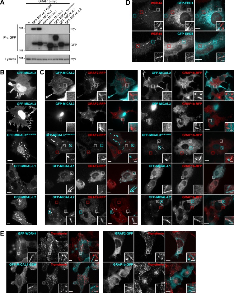
Figure S8.
Interplay of GRAF/WDR44 with other proteins of the MICAL family and with recycling endosomes. (A) Immunoprecipitation (IP) of transfected 293T cells with α-GFP. GRAF1b-myc was coimmunoprecipitated by GFP-MICAL1 and GFP–MICAL1 G3W but not by other members of the MICAL family. (B) Confocal stacks of transfected HeLa cells. As reported before, GFP-MICAL2 and GFP-MICAL3 were mostly nuclear but were also found at the plasma membrane (Giridharan and Caplan, 2014). A longer isoform of MICAL3, GFP-MICAL3pF1KA0819, was also nuclear but labeled thick and relatively static cytoplasmic tubular structures (Grigoriev et al., 2011). GFP–MICAL-L1 localized to intracellular tubules and puncta. GFP–MICAL-L2 was found at the plasma membrane and on intracellular puncta and tubules. (C) Confocal images of transfected HeLa cells. GFP-MICAL2, GFP-MICAL3, and GFP–MICAL-L1 did not colocalize with GRAF1b/2–RFP. GFP-MICAL3pF1KA0819 and GFP–MICAL-L2 displayed partial colocalization with GRAF1b/2–RFP tubules. Red boxed areas correspond to GRAF1b/2 tubules devoid of MICAL3pF1KA0819/MICAL-L2; cyan boxed areas correspond to MICAL3pF1KA0819/MICAL-L2 structures devoid of GRAF1b/2; white boxed areas show regions of colocalization and are magnified. Upon cotransfection of GFP–MICAL-L1, GRAF1b/2–RFP were essentially cytosolic and not found on tubules. (D) Confocal images of transfected HeLa cells stained with α-WDR44. Boxed areas show tubules positive only for WDR44 (red), only for GFP–EHD1/3 (cyan), or shared by WDR44 and GFP–EHD1/3 (white). (E) Confocal images of transfected HeLa cells incubated with Alexa Fluor 546–Transferrin (10 µg/ml, 30 min). GFP-tagged WDR44, MICAL1 G3W, GRAF2, or GRAF1b did not colocalize with Transferrin-positive endosomes. (B–E) Insets show magnifications of the boxed areas. Scale bars: 10 µm; scale bars of insets: 2 µm.

