Figure 5.
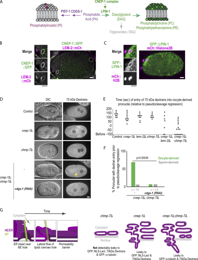
Spatial control of glycerolipid flux promotes nuclear closure. (A) Schematic showing de novo glycerolipid synthesis pathway in metazoans. (B and C) Confocal image of oocyte from time lapse series of oocyte-derived pronucleus expressing CNEP-1::GFP and LEM-2::mCherry in B or GFP::LPIN-1 and mCherry::Histone2B in C. (D) DIC and fluorescence images of embryos from adult worms injected with 70 kD Texas Red dextrans. (E) Plot of nuclear entry of 70-kD dextrans in oocyte-derived pronucleus for the indicated conditions. (F) Percent of pronuclei with dextran entry before pseudocleavage regression for the indicated conditions. P value reported is from a χ2 test. Scale bars, 5 µm. (G) Left: Working model for narrowing of NE holes by lateral flow of ER membrane sheets to restrict passage of large macromolecules. Right: Schematic model of defects in nuclear hole closure in different genetic backgrounds investigated in this study.

