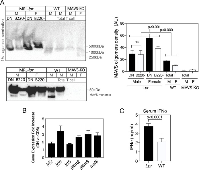
Figure 2.
MRL-lpr CD4–CD8– TCR-αβ+ cells manifest spontaneously oligomerised mitochondrial antiviral stimulator (MAVS) and increased expression of type I interferon (IFN-I)-stimulated genes (ISG) and serum IFN-I. (A) Lymph node cells from MRL-lpr mice were separated into CD4–CD8– TCR-αβ+ (double-negative (DN)) and CD4+ plus CD8+ (B220−) subsets and cell lysates prepared. Comparisons were made with lymph node cells from MRL+/+ wild-type (WT) or MAVS-knockout (KO) mice. Lysates were subjected to 1% agarose gels (upper panel) to selectively visualise MAVS oligomers, or SDS-PAGE (sodium dodecyl sulfate–polyacrylamide gel electrophoresis; lower panel) to visualise total MAVS monomer levels. (B) Fold increase in RNA expression for several ISG comparing female MRL-lpr DN to naive CD8+CD44low cells of the same mice. RNA was hybridised to Affymetrix GeneChip Mouse 430 2.0. Results are from five untreated 10-week-old mice per experiment and performed three times. (C) Serum levels of IFN-I in female MRL+/+ WT and MRL-lpr mice. Shown are the mean±SEM of five mice of each type. Statistical analysis was by unpaired t-test. Results were very similar in three experiments. F, female; M, male.

