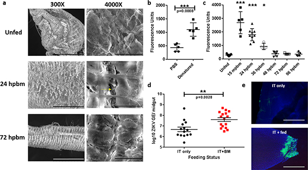
Figure 2: Bloodmeal acquisition induces microperforations in the midgut basal lamina.
(a) Representative SEM images of naïve and engorged Aedes aegypti midguts 24 hpbm and 72 hpbm at 300x (scale bars, 300 μm) and 4000x (scale bar, 20 μm) magnification. Two experimental replicates with 5 midguts/ replicate. Black dashed lines outline the visceral musculature and the yellow arrow denotes microperforations in the underlying basal lamina. (b) Pools of 5 unfed Ae. aegypti midguts per replicate were assayed for fluorescein labeled collagen hybridizing peptide (CHP) binding (n=5). Samples were analyzed by two-tailed t-test (p=0.0008). (c) Temporal binding profile of CHP binding to pools of 5 midguts/ replicate following a single bloodmeal. Differences in CHP binding were determined using a One-way ANOVA with a Dunnett’s multiple comparisons post-test. Precise p-values and samples sizes can be found in the linked source data. (d) Viral genome equivalents/ midgut of individuals intrathoracically inoculated with ZIKV (n=14) or inoculated and provided a bloodmeal 3 dpi (n=16). Midguts were assayed 12 dpi by RT-qPCR and statistically analyzed with a two-tailed t-test. (e) Midguts from ZIKV inoculated individuals with or without a bloodmeal assayed for ZIKV antigen 12 dpi. Representative images of two experimental replicates of 10 individual midguts/ per group. Images were acquired at 200X magnification (scale bar, 100 μm). For b-e, center values represent the means and error bars represent the SD.

