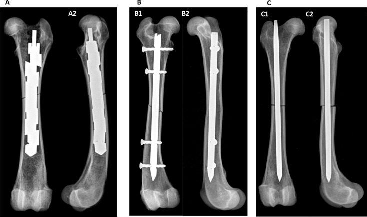
Fig 2.
Radiographs of canine femora with implanted (A) EXPN, (B) ILN and (C) STMN. (A) Caudocranial (A1) and mediolateral (A2) radiograph of a femur with an oblique fracture and implanted and expanded EXPN. (B) Caudocranial (B1) and mediolateral (B2) radiograph of a femur with oblique fracture and implanted ILN.

