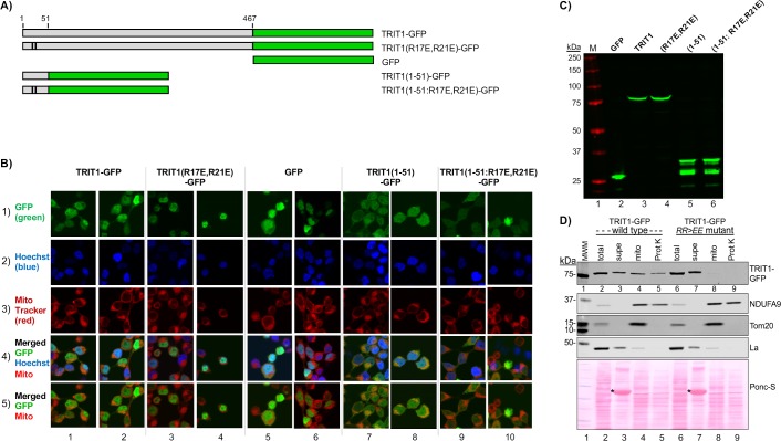
Fig 2. Human TRIT1 contains an N-terminal mitochondrial targeting sequence (MTS).
A) Cartoon of the GFP-fusion constructs. Numbering represents amino acids of TRIT1; black vertical lines indicate positions of two mutated residues in the R17E/R21E constructs. B) Representative confocal microscopic images from HEK293 cells transfected 48 hours prior with the constructs in A and stained with Hoechst and MitoTracker. C) Western blot from cells transfected with constructs in A) using anti-GFP antibody (Ab). D) Western blot subcellular fractionation analysis. Transfected HEK293 cells were fractionated 48-hour later into supe (cytosol), mitochondria (mito) and mitochondria treated with Proteinase K (ProtK), the latter to digest proteins on the outer membrane of the mitochondria. GFP-TRIT1 WT and RR>EE mutant was detected using anti-GFP Ab. NDUFA9 is a mitochondrial inner membrane protein, La is a nuclear and cytosolic protein, TOMM20 is on the mitochondrial outer membrane. Total protein staining with Ponceau S is shown in the lower panel. Asterisks indicate BSA that was added during the mitochondrial isolation protocol (see text).

