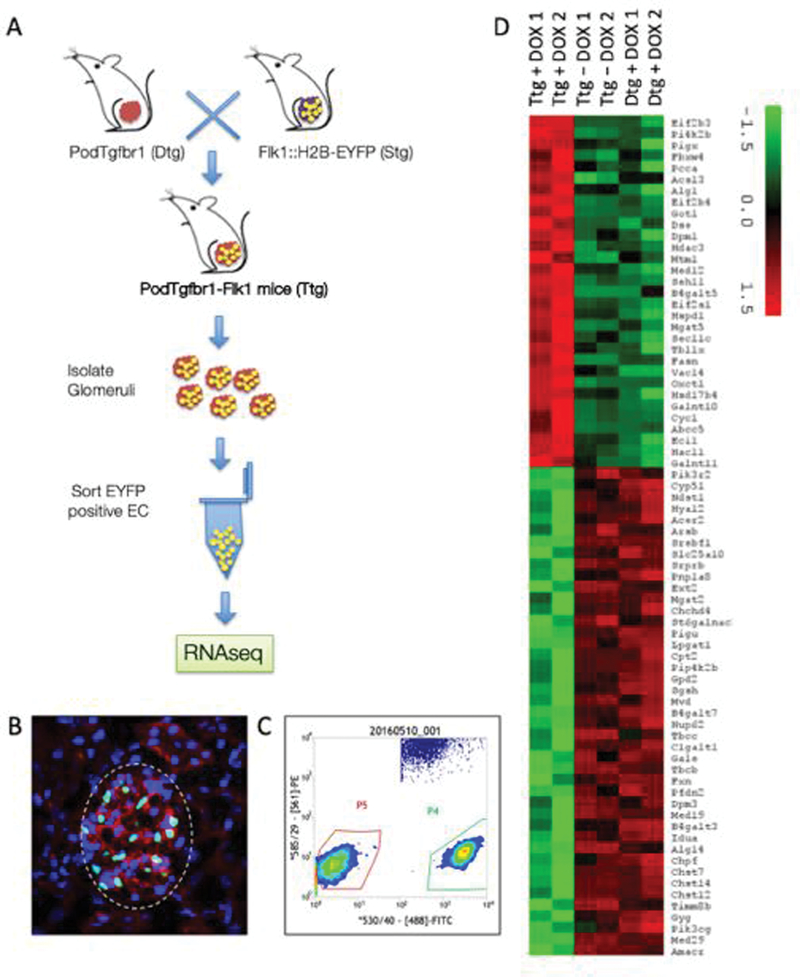
Figure 2. Tgfbr1 signaling in podocytes of Flk1::H2B-EYFP mice results in activation of endothelial surface layer (ESL) degradation pathways.
(A) Mating strategy of PodTgfbr1 double transgenic (Dtg) (FVB background) crossed with Flk1::H2B-EYFP mice (10 generation backcrossed to FVB) to generate PodTgfbr1-Flk1 mice with GECs expressing enhanced yellow fluorescent protein (EYFP) positive nuclei. Pod-Flk1 (Dtg) and PodTgfbr1-Flk1 (Ttg) mice were fed +/− DOX and glomeruli we isolated after 4d of treatment and followed by FACS sorting of EYFP cells. (B) Representative glomeruli from PodTgfbr1-Flk1 Ttg mice showing DAPI (blue) and EYFP positive GEC nuclei (green) and CD31 cellular staining (red). (C) EYFP positive GECs were sorted and isolated by FACS (Gated P4) from controls and 4 days of DOX treated PodTgfbr1-Flk1 mice as described in 58. The Gated P5 cells were negative EYFP glomerular cells. 10,000–30,000 EYFP positive GECs/mouse were collected and 3–6 mice were pooled to yield sufficient RNA for sequencing per sample run. Transcriptomic assessment of GECs isolated from Flk1::H2B-EYFP mice. (D) Heatmap of genes in glycosaminoglycan related pathways show the differential expression of genes in these pathways between triple transgenic (Ttg) PodTgfbr1-Flk1 mice + day 4 DOX, double transgenic (Dtg) Pod-Flk1 mice + day 4 DOX, Ttg PodTgfbr1-Flk1 mice no -DOX control mice.

