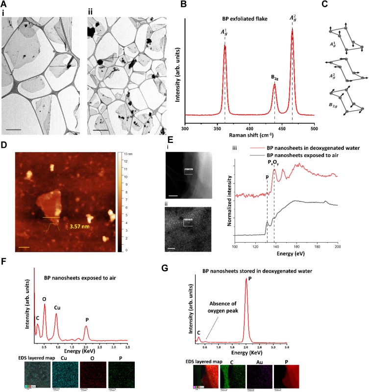
Figure 2.
Morphological characterization and elemental analysis of BP nanosheets. (A) Low-magnification TEM images of exfoliated BP ultra-large nanosheets. In image i, the scale bar represents 1 µm, and in image ii, the scale bar is 2 µm. (B) Raman spectra of exfoliated BP showing characteristic peaks of BP. (C) Representative schematic of out-of-plane and in-plane vibrational modes for BP – Raman analysis. (D) AFM analysis to confirm the exfoliated BP nanosheets thickness variation (scale bar is 50 nm). (E) EELS analysis: i. The high-angle annular dark-field (HAADF)-STEM image of BP flake exposed to air, acquired at 80 kV (scale bar is 20 nm). ii. The HAADF-STEM image of BP flake in deoxygenated water (scale bar is 0.2 µm). iii. The EELS spectra corresponding to the selected area in i and ii shows the P-L2,3 edge of bulk BP and BP nanosheet confirming the BP nanosheets stored in the deoxygenated water were not oxidized. (F) The EDS analysis of BP flake exposed to air along with elemental mapping confirming the presence of oxygen (scale bar is 25 nm). (G) The EDS analysis of BP flake stored in deoxygenated water along with elemental mapping confirming the absence of oxygen (scale bar is 250 nm).

