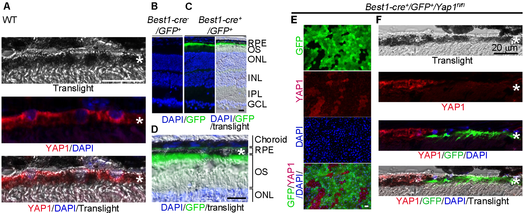
Figure 1.

Mosaic deletion pattern of Yap1 mediated by Best1-Cre in adult RPE in vivo. A, Immunostaining of YAP1 showed cytoplasmic localization in the RPE cells of wild-type mouse at 6-week-old. B,C, Best1-Cre was confirmed by GFP expression in the RPE of Best1-Cre+/GFP+ (C) but not Best1-Cre−/GFP+ mice (B). D, High magnification of image of the right panel in (C). E,F, YAP1 immunostaining showed that YAP1 was only depleted in GFP+, but not in the GFP− RPEs of the Yap1 cKO mice (Best1-Cre+/GFP+/Yap1fl/fl, MUT). RPE whole mount (E) and RPE tissue section (F) from the Yap1 cKO mice were immunostained by YAP1 (red), GFP (green), and DAPI (blue). *RPE layer. Bar size = 20 μm. At least 5 animals per group were used for each assay.
