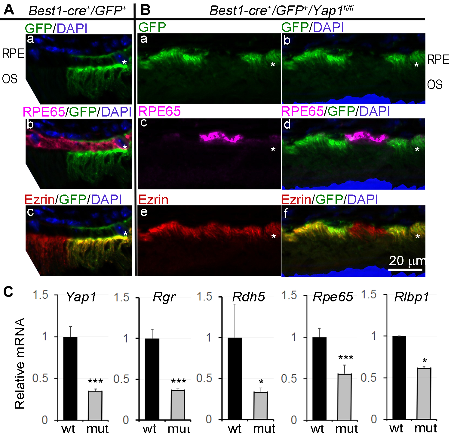
Figure 3.

Loss of RPE65 expression in Yap1 mutant RPE cells. Yap1 cKO mice (Best1-Cre+/GFP+/Yap1fl/fl, MUT) and the control mice (Best1-Cre+/GFP+, WT) were analyzed at 5–8 weeks of age. A,B, Immunostaining of RPE differentiation markers, RPE65 and Ezrin on the WT (A) and mutant (B) retina sections of 5-week-old mice. * in (A) and (B): RPE layer. Bar size = 20 μm. n = 5 mice for each group. C, qPCR analysis showed reduced expression of RPE65 and other visual cycle genes. Value = mean ± SD. n = 3 samples for each group. Each sample was extracted from pooled RPE sheets of 5 mice. *, *** are P < .05, P < .005, respectively.
