
Figure 2.
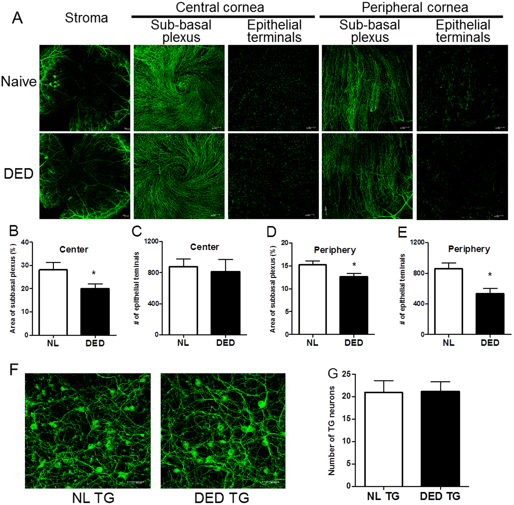
Morphological changes in corneal innervation after desiccating stress. (A) Representative images of corneal whole mount with β-tubulin III immunostaining showed stromal nerves, subbasal plexus and epithelial terminals in the central and peripheral cornea in naïve and mice with DED (n=10 per group). Stromal nerve: x50 magnification. Subbasal plexus and epithelial terminals: x200 magnification. (B-E) Density of subbasal plexus was shown as area occupied by nerve fibers (%) per microscope field (B &D). Density of epithelial terminals was shown as number of terminals per microscope field (C&E). (F) Representative images of cultured TG neurons (β-tubulin III; green) isolated from naïve (n=5) and mice with DED (n=5), x400 magnification. (G) Number of TG neurons counted per microscope field was shown. Error bars represent means ± SEM. *p<0.05.