
Figure 5.
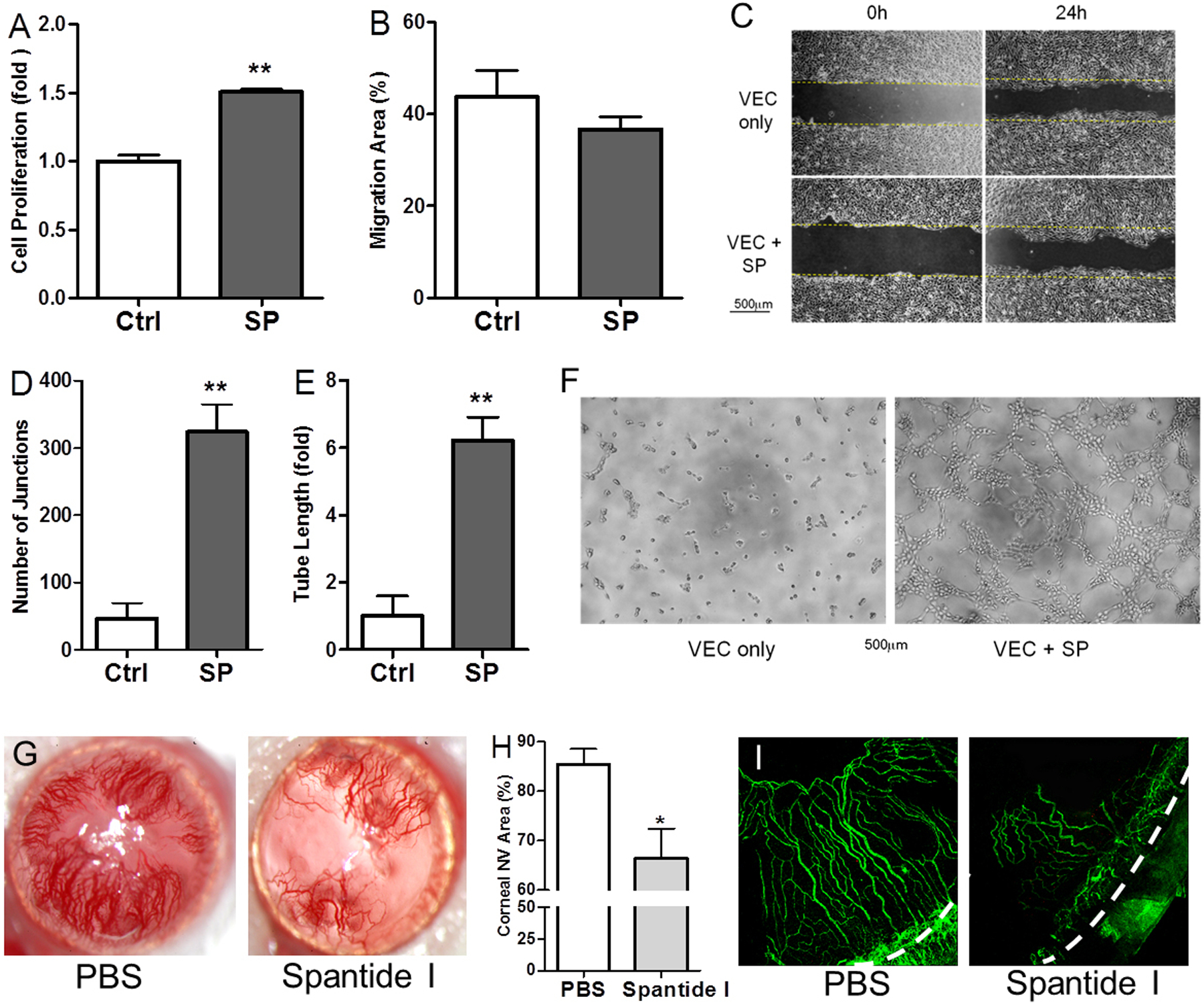
SP promotes angiogenesis in vitro and in vivo. (A) VEC proliferation was determined after 24-hour culture with SP, n=6. (B-C) Migration of VEC when cultured with SP was quantified (B) and photographed at 0h and 24h (C), n=5. (D-F) Tube structure formation in the matrigel cultured with SP was photographed at 4h (F) and quantified as the number of junctions (D) and length of tube formed (E), n=4. Scale bars, 500μm. (G) Representative images of corneal NV with slit lamp photography. (H) Quantification of corneal NV area (%) of slit lamp photography at 14 days after vehicle and spantide I treatment (n=10 per group). (I) Immunohistochemistry with CD31 to highlight corneal NV, x100 magnification, dashed line indicates limbus. *p<0.05, **p<0.01.