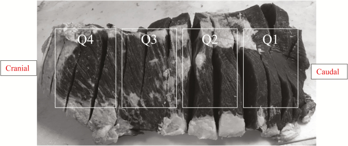
Figure 1.
Quadrants of the outside round where injection-site lesions were evaluated. Q1 was identified as the most caudal end and was the closest to the shank, whereas Q4 was identified as the most cranial end and only included the biceps femoris muscle. Q2 and Q3 were evenly split between Q1 and Q4. Q1, Q2, and Q3 included both the biceps femoris and the semitendinosus muscles.

