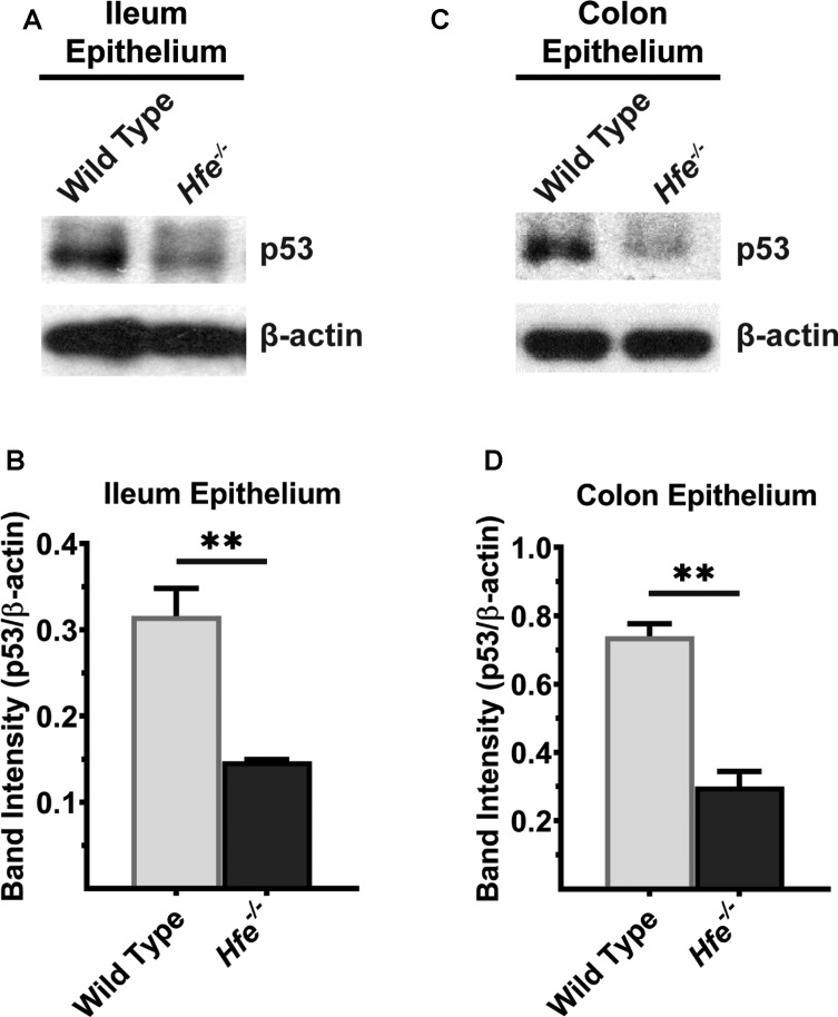
Figure 7. p53 protein levels are significantly decreased in Hfe−/− mouse ileum and colonic epithelium.
Western blot for p53 protein in (A) ileal and (C) colonic epithelium of wild type and Hfe−/− mice. Please see Supplementary Figures S13 and S14 for full blot images. Western blot band intensities were estimated using ImageJ software and the band intensities that correspond to p53 protein were normalized to β-actin levels in (B) ileal epithelium and (D) colonic epithelium. Data represent mean values of three mice per group ± SEM. **P < 0.01.

