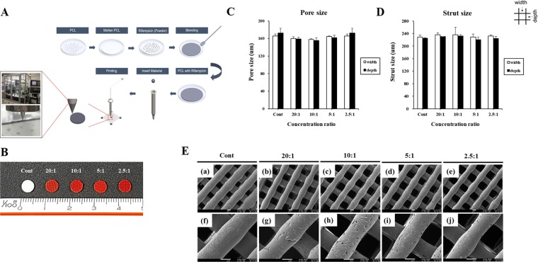
Figure 1.
Fabrication of a rifampicin-loaded 3D scaffold. (A) Schematic illustration of the procedure involved in the fabrication of the rifampicin-loaded scaffold using our 3D printing system. (B) Photographs of the rifampicin-loaded scaffold at different concentrations.. Electron micrographs were analyzed using microscope to determine the (C) pore size and (D) strut size of the scaffolds. (E) SEM image of scaffolds with rifampicin concentration of (a,f) Cont, (b,g) 20:1, (c,h) 10:1, (d,i) 5:1, and (e,j) 2.5:1. The image of (f–j) indicate magnified view of images of (a–e), respectively. a–e: x70, f–j: x200, scale bar: 100 um.

