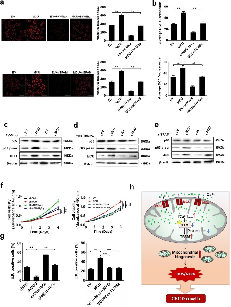
Fig. 7.
Mitochondrial Ca2+-mediated mitochondrial biogenesis promotes CRC proliferation by ROS/NF-kB signaling. a Immunofluorescence images (Left) of mitochondrial ROS (mROS) and MitoSOX fluorescence intensity (Right) in LS174T cells, treated as indicated. b Average DCF fluorescence intensity in LS174T cells treated as indicated. Western blotting analysis to measure the expression levels of p65 and pi-p65 in LS17T cells with MCU overexpression or treated with c PV-Mito, d Mito-TEMPO, or e siTFAM. f MTS assay to measure cell viability and g EdU incorporation assays to measure cell proliferation in LS174T cells, treated as indicated. h Schematic representation showing the underlying mechanism of MCU-mediated mitochondrial Ca2+ uptake in the promotion of CRC growth. *P < 0.05; **P < 0.01. MCU mitochondrial calcium uniporter, CRC colorectal cancer, TFAM transcription factor A, mitochondrial, ROS reactive oxygen species, PV-Mito expression vector encoding parvalbumin with mitochondria target sequence, si small interfering, Ca2+ calcium

