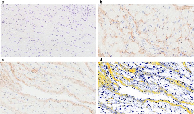
Figure 5.
Tetranectin immunohistochemical tissue staining. Human cardiac tissue was obtained from cardiac by-pass patients, formalin-fixed and paraffin-embedded for immunostaining with either a rabbit anti-human monoclonal antibody against Tetranectin or an IgG isotype control antibody. Sections were counterstained with Haematoxylin and imaged using Aperio ScanScope digital scanner. A positive pixel count algorithm was applied to analyse the area and intensity of immunoreactivity. Representative images presented; IgG control (a); examples of Tetranectin (brown) and haematoxylin (blue) stained tissue sections (b,c); Example of the Tetranectin positive pixel algorithm used to quantify tissue protein expression (d). Images were captured at 20x magnification.

