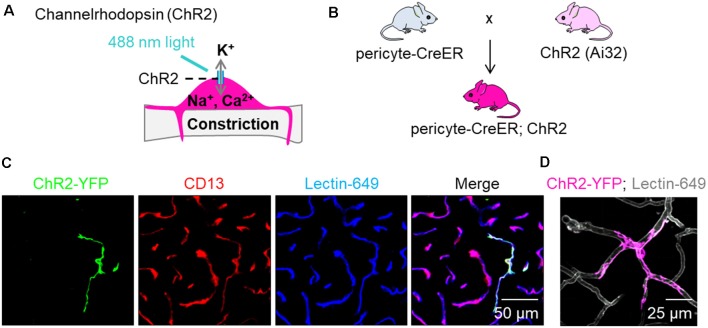
Figure 1.
Expression of channelrhodopsin (ChR2) in pericytes. (A) Schematic demonstrating our hypothesis that excitation of ChR2 expressing pericytes by 488 nm light will depolarize pericytes causing them to contract and constrict the underlying capillary. (B) Schematic showing the breeding scheme of recently characterized double promoter pericyte-CreER mice with tamoxifen inducible Cre-recombinase expression in pericytes, with mice that have a loxP-flanked STOP cassette that is excised in the presence of Cre to drive ChR2-EYFP fusion protein expression in pericytes (Ai32, Jackson Laboratory); the crossed mice were termed pericyte-CreER; ChR2. (C) Representative images of EYFP expression (green) showing co-localization of ChR2 cation channels with CD13+ pericytes (red) on Lectin-649+ capillary profiles (blue) in pericyte-CreER; ChR2 mice treated with tamoxifen (40 mg/kg i.p. daily for 4 days) and studied 2 weeks after the last tamoxifen injection. (D) In vivo z-stack maximum projection image demonstrating EYFP (pink) in a pericyte along lectin-649 + (gray) capillary profiles in a 32-month-old female pericyte-CreER; ChR2 mouse.

