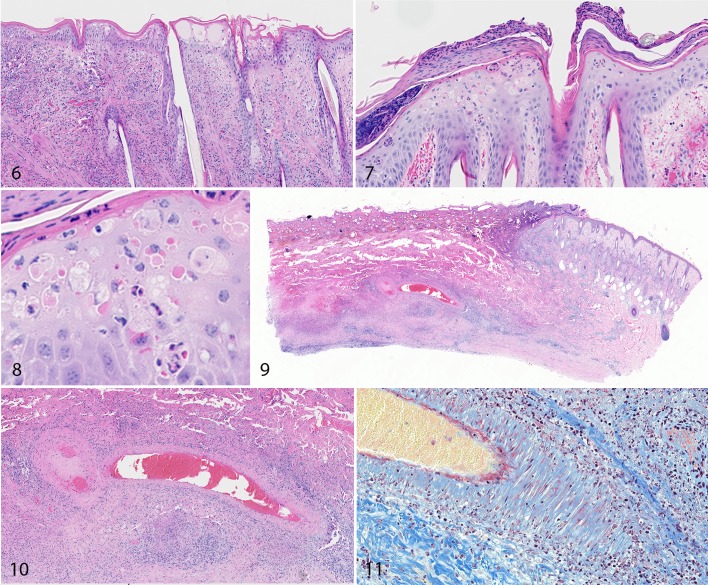
Figures 6–8. Acute lumpy skin disease, skin, calf, 9 days post inoculation (DPI; Fig. 6) and 15 DPI (Figures 7, 8). Degeneration and necrosis of keratinocytes, intracytoplasmic inclusion bodies, and vesicles are present in the epidermis. Edema, hemorrhage, and influx of lymphocytes and macrophages are present in the dermis. HE. Figures 9–11. Lumpy skin disease, skin, calf. There is a well-demarcated wedge-shaped region of necrosis (infarct; Fig. 9). In the center of the infarct, the wall of a large muscular blood vessel is disrupted by mononuclear inflammatory cells and fibrin (fibrinonecrotic vasculitis; Figure 10 and red staining in Figure 11). HE (Figs. 9, 10). Martius scarlet blue trichrome stain (Fig. 11).

An official website of the United States government
Here's how you know
Official websites use .gov
A
.gov website belongs to an official
government organization in the United States.
Secure .gov websites use HTTPS
A lock (
) or https:// means you've safely
connected to the .gov website. Share sensitive
information only on official, secure websites.
