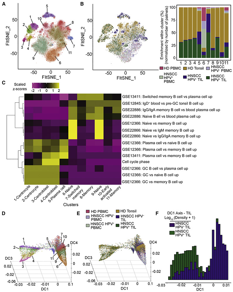
Figure 4. Analysis of tonsil and TIL B cells reveals granular details of germinal center B cells, and a unique B cell population associated with HPV− TIL.
A total of 16,736 B cells were recovered from all samples. (A-B) We identified a total of 11 clusters of B cell from tonsils, TIL and PBMC. (C) Gene set enrichment revealed a germinal center phenotype associated with clusters 1-4, enrichment of genes for plasma cells in cluster 5, and combinations of naïve, memory and switched B cells in other clusters. (D) Diffusion map embedding of all B cells colored by clusters as in (A). This three-dimensional embedding yielded axes related to germinal center formation (DC1), transition from naïve to memory B cells (DC4) and progression to plasma cells (DC3). Few HPV− B cells progress along DC1 to become germinal center B cells. (E) Same diffusion map embedding of as in (D), but colored by sample types. (F) The majority of HPV− B cells are concentrated on the right side of the DC1 axis, while HPV+ cells have a bimodal distribution along the DC1 axis (note log scale on the y axis in [F]).

