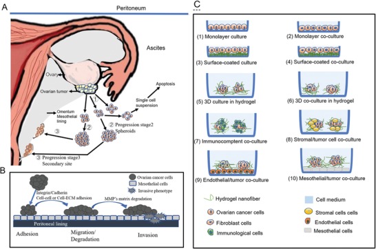
Figure 4.

Tumor progression, peritoneal dissemination, cancer cell behaviors in ovarian cancer and schematic cell culture patterning for precise oncology remodeling in vitro. A) Schematic multistep tumor progression and peritoneal dissemination in the abdominal cavity, ➀ Single EOC cells from primary ovarian cancer site on ovary surface; ➁The primary cancer cells aggregate to survive as tumor spheroids in the ascites‐rich TMEs; ➂Partially malignant cells metastasize to the secondary site and interact with mesothelial cells to form new metastasis on the omentum. B) A schematic diagram of early‐stage primary cancer cell behaviors within a normal epithelium with breach of the peritoneal lining that separate it from the underlying stromal tissue, such as adhesion mediated by integrin/cadherin, migration promoted by extracellular solutes (growth factors, cytokines, and chemokines), and invasion induced by enzyme degradation. C) Schematic cell patterning culture models in vitro. The cartoon diagram displays the main in vitro 2D and 3D culture or coculture models, that are used to study intercellular and cell‐TME crosstalk. Two or three types of cells are incorporated to the schematic spherical 3D coculture cell models or other cell patterns.
