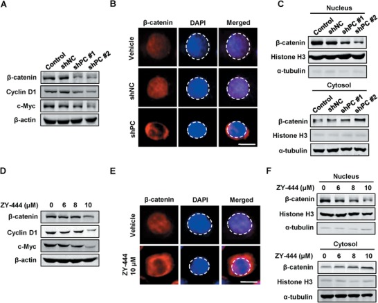
Figure 4.

Both the deletion of PC and ZY‐444 treatment inhibited the Wnt signaling pathway in MDA‐MB‐231 cells. A) The expression of key downstream proteins of the Wnt signaling pathway in control cells, shNC, shPC #1, and shPC #2 cells. B) The representative immunofluorescent images for control, shNC, and shPC MDA‐MB‐231 cells for β‐catenin (red) and DAPI (blue) signals. The DAPI signal was circled using dotted lines. Scale bar = 5 µm. C) The protein expression of β‐catenin and Histone H3 in nuclear and cytosolic fractions of control, shNC, and shPC MDA‐MB‐231 cells. D) The expression of downstream proteins of the Wnt signaling pathway in MDA‐MB‐231 cells under ZY‐444 treatment. E) Representative images of stained MDA‐MB‐231 cells treated with vehicle or ZY‐444. The red and blue staining represent β‐catenin and nucleus, respectively. The nucleus was circled using a dotted line. Scale bar = 5 µm. F) Effects of ZY‐444 on β‐catenin distribution between nuclear and cytosolic components in MDA‐MB‐231 cells.
