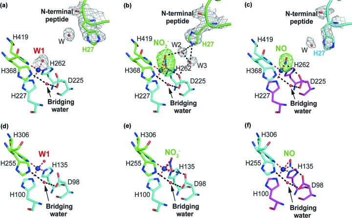
Figure 3.
Ligand-bound structures of Hd 1NES1NiR compared with those of AcNiR. (a) Ligand water (W1)-, (b) nitrite (NO2 −)- and (c) nitric oxide (NO)-bound T2Cu of Hd 1NES1NiR colored green, magenta and cyan for each monomer. The T2Cu ion is represented by a deep-blue sphere. The water molecules are represented by red spheres and the bridging water is indicated by a black arrow. Coordination to the T2Cu ion is shown by a red broken line and the interaction is shown by a black broken line. The F o F c electron density map at the 5.0σ level is shown for nitrite (NO2 −) and nitric oxide (NO). The 2F o F c electron-density map at the 1.0σ level is shown for the His27, the ligand-water (W1) and the other waters (W2, W3, W). (d) The ligand water (W1)-, (e) nitrite (NO2 −)- and (f) nitric oxide (NO)-bound T2Cu of the AcNiR are colored green and cyan for each monomer. The T2Cu ion is represented by a deep-blue sphere. The water molecules are represented by red spheres and the bridging water is indicated by a black arrow. Coordination to the T2Cu ion is shown by a red broken line and the interaction is shown by a black broken line. The structural coordinates for (d), (e) and (f) are from the PDB entries 6gsq and 6gto (Halsted et al., 2019 ▸), and 5of8 (Horrell et al., 2018 ▸), respectively.

