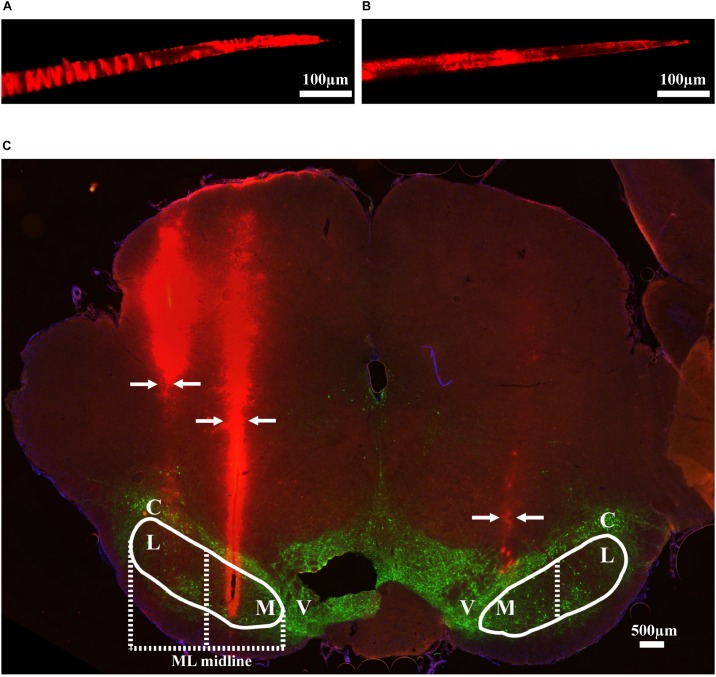
FIGURE 2.
Methods used to validate electrode location. (A) An example of DiI coated electrode before brain insertion. (B) The same electrode after brain insertion. (C) Coronal brain section showing DiI coated electrodes in mSNr and lSNr. Red indicates electrode tracks coated with DiI; green indicates dopaminergic neurons in SNc and VTA stained by anti-tyrosine hydroxylase. Borders of SNr are indicated by the white solid line. mSNr and lSNr (separated by dashed white line) were defined by evenly dividing the SNr in the medial/lateral direction. Arrows indicate locations of the electrode tracks. Abbreviations: M, medial Substantia Nigra pars reticulata (mSNr); L, lateral Substantia Nigra pars reticulata (lSNr); C, Substantia Nigra pars compacta (SNc); V, ventral tegmental area (VTA).

