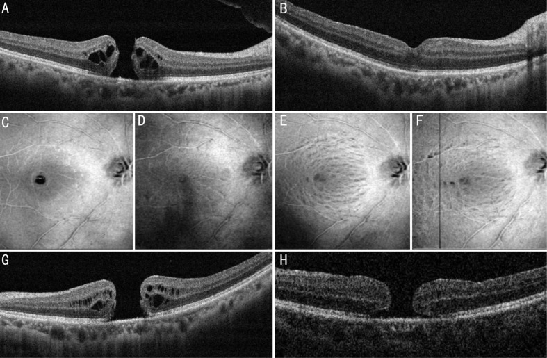
Figure 3. Results of the surgery.
A, B: A 64-year old male presented with an MH of 365 µm. A: The intraoperative ILM-retina adhesive force was graded mild; B: Three months later, the MH showed a good configuration and the BCVA improved from 20/100 preoperatively to 20/32 postoperatively. C-F: 3D wide-field en-face scans of SD-OCT showing the ILM layer. C: Before the surgery; D: One month after the surgery, the MH was closed and no severe iatrogenic damage caused by the modified technique was detected; E: Three months after the surgery; F: One year after the surgery. Inner retinal dimplings were observed in the ILM-peeled area, which is similar to other patients who underwent conventional ILM peeling. G, H: A 47-year old female with an MH of 659 µm. G: The intraoperative ILM-retina bonding force was graded mild; H: After one-month follow-up, the MH remained unclosed with a smaller diameter of 540 µm. She received another lens capsular flap transplantation and the MH was closed.

