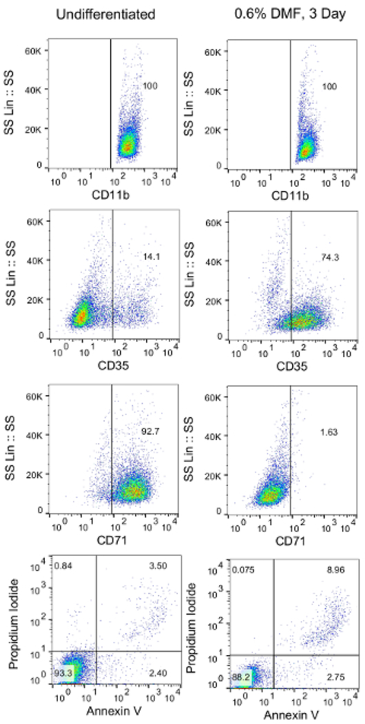
Figure 1: Validation of HL-60 cell differentiation via flow cytometry.
Differentiated HL-60 cells were harvested, washed, and resuspended in 1 × 10⁵ cells/mL PBS. Cells were then aliquoted into 12 wells (100 μL/well) in a 96-well plate. Cells were then stained with fluorescently conjugated anti-CD35, anti-CD71, annexin V, and propidium iodide. Unstained cells or cells stained with fluorescently conjugated isotype antibodies were used as controls.

