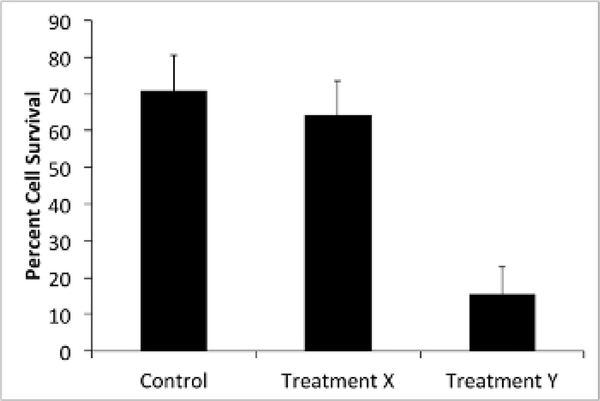
Figure 2: Treatment Y improves HL-60-mediated cell killing of bacteria.
S. pneumoniae samples were treated with treatment X (antibody) or treatment Y (enzyme). OPKA was performed according to the protocol and bacterial CFUs were counted in duplicate. Samples that were not treated with HL-60 cells were used as a control (100% cell survival). Shown are the average percentages of bacterial CFUs in the HL-60 treated groups compared to the corresponding non-HL-60-treated groups. Bars represent standard error.

