Figure 1.

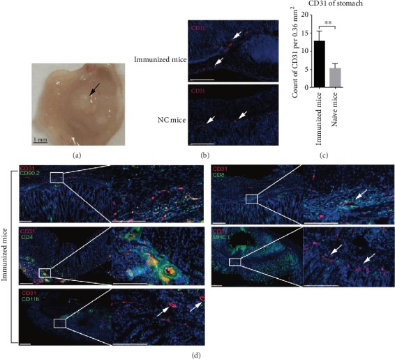
Formation of perivascular infiltrates of CD4/CD8+ T clusters in inflammation-experienced gastric subserous tissue. Six to eight-week-old female C57BL/6 mice were vaccinated in the gastric subserous layer. (a) 28 days later, the gastric tissue was exposed and a white lump was observed. (b, c) Frozen sections of gastric tissue from immunized or naïve mice were stained with antibodies against CD31 (red); nuclei are depicted by DAPI stained blue. Scale bars: 100 μm. The fluorescence images were acquired with a Panoramic 250 Flash III Scanner (3DHistech); counts of CD31 per 0.36 mm2 were randomly chosen and calculated. Values are the mean ± SD (n = 3). ∗∗P < 0.01. (d) Frozen sections of gastric tissue from immunized mice were stained with antibodies against CD31 (red), CD90.2, CD4, CD8, CD11b, or MHCII (green); nuclei were depicted by DAPI stained blue. Scale bars: 200 μm.
