Figure 4.

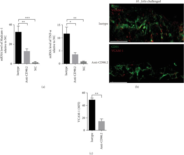
Perivascular lymphocyte clusters might increase the expression of VCAM-1, Madcam-1, and TNF-α. Mice were treated as indicated in Figure 2(a). (a) The expression level of Madcam-1 and TNF-α in the gastric tissue of the indicated partner mice challenged with H. felis was analyzed by qRT-PCR. (b) Frozen sections of gastric tissue from the indicated partner challenged with H. felis were stained with antibodies against CD31 (green) and VCAM-1 (red), and nuclei were treated with DAPI (blue). Scale bars indicated 50 μm. (c) Mean fluorescence intensity (MFI) of VCAM-1 expression on CD31+ vessels was determined. Values are the mean ± SD (n = 3). All indicated P values were tested using the ANOVA analyses or t tests. ∗∗P < 0.01, ∗∗∗P < 0.001, and ∗∗∗∗P < 0.0001; n.s.: not significant.
