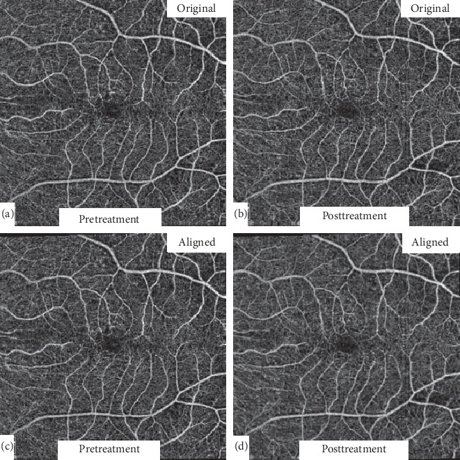
Figure 1.

Automated image alignment. Original 6 × 6 mm pre- and posttreatment images (a and b) were automatically registered and aligned (c and d) using a commercially available retina alignment software to allow comparison of only areas common to both images.
