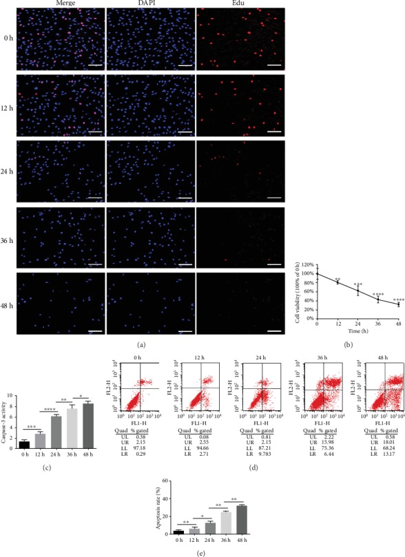
Figure 1.

Compression decreased cell viability and induced apoptosis in human NPMSCs. (a) The outcomes of the Edu-staining proliferation assay showed that NPMSC proliferation was progressively impaired by compression (scale bar = 100 μm). (b) The results of the CCK-8 assay suggested that NPMSC cell viability decreased in a compression time-dependent manner. Data were presented as the means ± SD (n = 3) (∗P < 0.05, ∗∗P < 0.01, ∗∗∗P < 0.001, and ∗∗∗∗P < 0.0001 compared with 0 h). (c) Quantitative analysis suggested increasing caspase-3 activity as compression prolonged. (d) Annexin-V/PI staining was performed to determine the apoptosis rate of human NPMSCs by flow cytometry; Annexin-/PI- represents live cells, Annexin+/PI- represents apoptotic cells on early stage, Annexin+/PI+ represents apoptotic cells on late stage, and Annexin-/PI+ represents necrotic cells. (e) Histogram analysis showed the percentage of apoptotic human NPMSCs (Annexin+/PI- plus Annexin+/PI+). Data were presented as the means ± SD (n = 3) (∗P < 0.05, ∗∗P < 0.01, ∗∗∗P < 0.001, and ∗∗∗∗P < 0.0001 between two groups). NPMSCs: nucleus pulposus mesenchymal stem cells; PI: propidium iodide; Edu: 5-ethynyl-2′-deoxyuridine.
