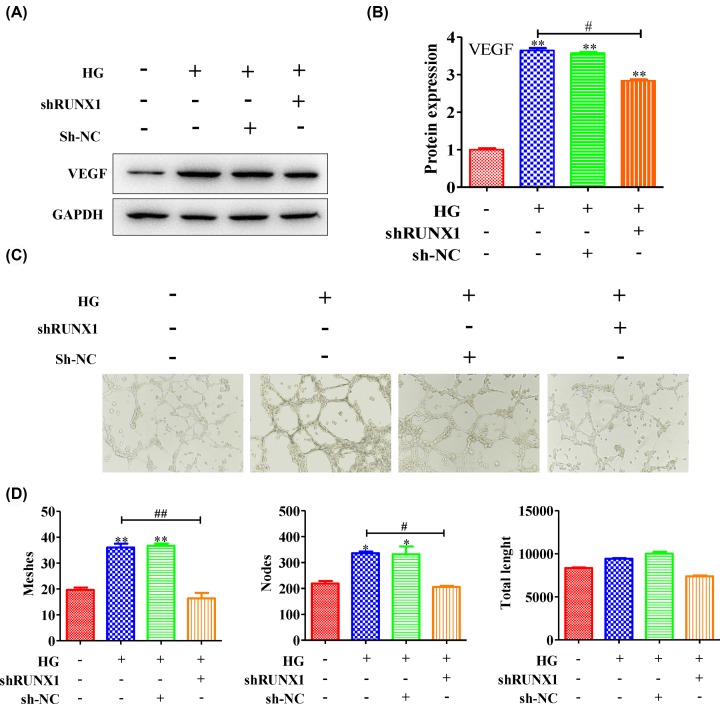
Figure 2. The angiogenesis level was inhibited by silencing RUNX1.
HRMECs were stimulated with or without high levels of glucose and then treated with or without RUNX1 adenovirus. (A) The expression level of VEGF was measured, and (B) the quantification was showed. Meanwhile, (C) the tubes that formed were photographed. (D) The number of tubes, nodes and the tube length were determined. All experiments were performed triplicate. Data are presented as the mean ± standard deviation from triplicate wells; *P<0.05 and **P<0.01 compared with the control; #P<0.05 and ##P<0.01 compared with HG-induced HRMEC cell group.

