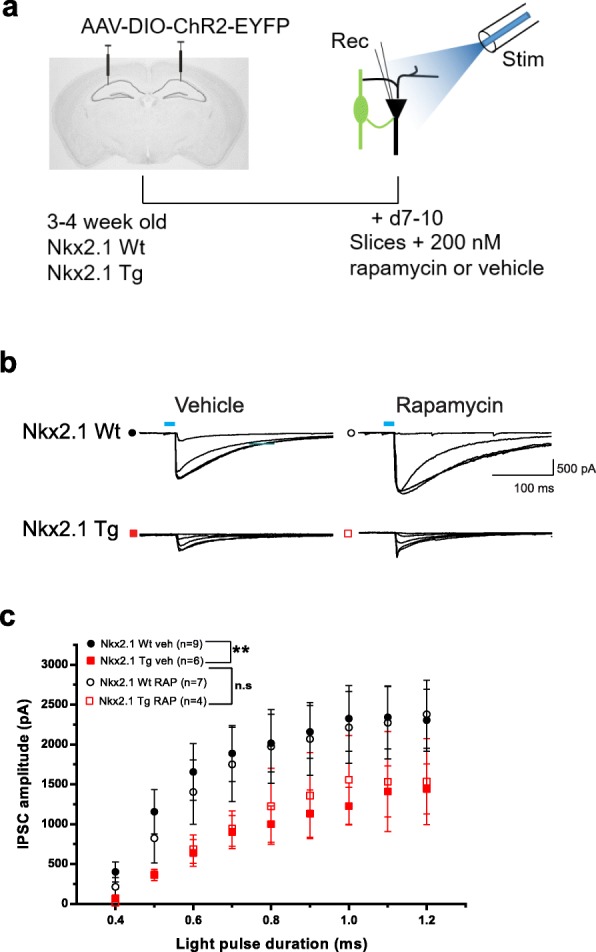
Fig. 6.

Acute rapamycin treatment did not affect the deficit in synaptic inhibition. a Diagram of experimental paradigm for acute treatment of slices with 200 nM rapamycin (or vehicle) and recording of synaptic inhibition of pyramidal cells by Nkx2.1 interneurons from Nkx2.1Cre/wt;Tsc1f/wt mutant and Nkx2.1Cre/wt control mice. b, c Representative traces (b) and summary graph (c) of input-output function of IPSCs evoked in CA1 pyramidal cells by optogenetic stimulation of Nkx2.1 interneurons in slices from Nkx2.1Cre/wt;Tsc1f/wt mutant (red) and Nkx2.1Cre/wt control (black) mice that received vehicle (filled symbols) or rapamycin (open symbols). Acute rapamycin treatment did not affect the reduction of IPSC amplitude in mutant relative to control mice (n = 9 cells in 4 animals for vehicle-treated and 7 cells in 4 animals for rapamycin-treated Wt mice (Nkx2.1Cre/wt;Tsc1wt/wt); n = 6 cells in 4 animals for vehicle-treated and 4 cells in 3 animals for rapamycin-treated Tg mice (Nkx2.1Cre/wt;Tsc1f/wt); 3-way ANOVA, Bonferroni tests, **p < 0.01, n.s. not significant)
