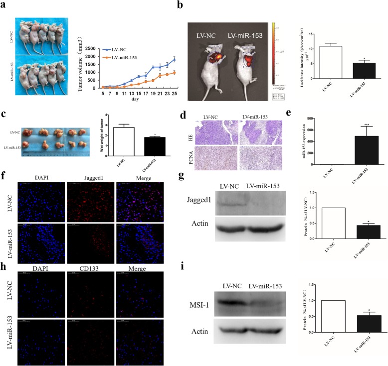
Fig. 4.
miR-153 overexpression inhibits tumor growth in vivo. a Subcutaneous tumor model of nude mice at 4 weeks after injection (n = 5 per group). Tumor sizes were measured every 2 days. b Representative image of subcutaneous tumor evaluated via in vivo bioluminescence assay at 25 days after injection. c The tumors were collected at 25 days after injection and the wet weight of tumor was measured by electronic scale. d HE staining and immunohistochemistry analysis of proliferating cell nuclear antigen (PCNA) in tumors from indicated SPC-A-1 cells bearing mice. Scale bar, 100 μm. e miR-153 expression in tumors from indicated SPC-A-1 cells bearing mice was determined by qPCR. f Jagged1 expression was determined by immunofluorescence. Scale bar, 50 μm. g Jagged1 expression was determined by Western blot. h Stem cell marker CD133 expression was determined by immunofluorescence. Scale bar, 50 μm. i MSI-1 expression was determined by Western blot. Values of Jagged1 or MSI-1 immunoreactivity derived from vehicle groups (LV-NC) was normalized to 1.0. Values of LV-miR-153 groups were normalized according to vehicle groups (LV-NC). Data shown are mean ± s.d. of three independent experiments. *P < 0.05 by two-tailed Student’s t test

