Fig. 1.
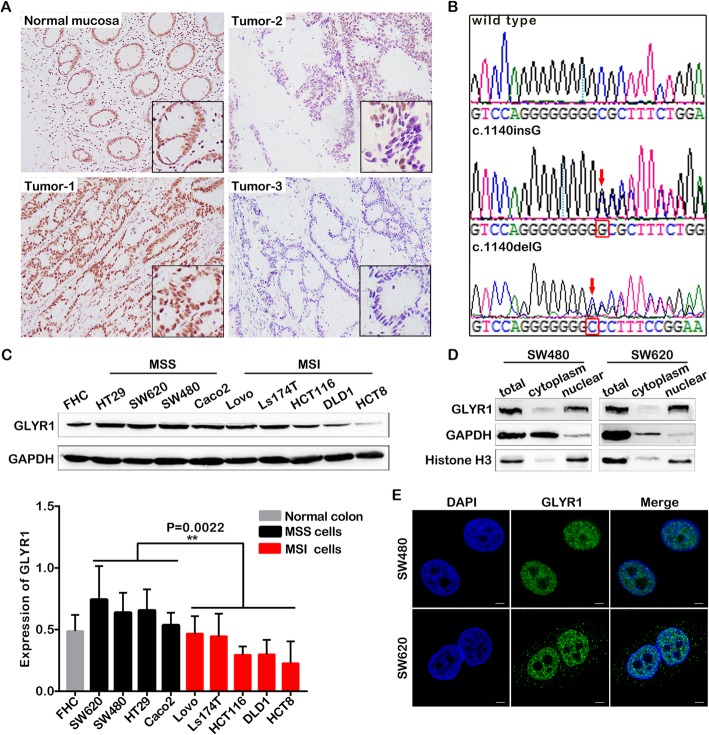
GLYR1 is downregulated in MSI CRC. a Representative photographs showing immunohistochemical staining of GLYR1 (× 100, scale = 100 μm) in 221 paraffin-embedded normal human colorectal tissue (normal mucosa) and colorectal cancer tissue (Tumor-1, Tumor-2, Tumor-3) sections. The image in the black pane in the lower right corner of the overlay shows a partial enlargement. b GLYR1 exon 13 mutation detection in CRC tissues and cells. Partially intercepted representative GLYR1 exon 13 normal (wild type) and mutant (c.1140insG, c.1140delG) sequences are shown. The red arrowhead indicates the mutation site. c Western blot analysis of GLYR1 protein expression in CRC cells. Quantification of protein expression was normalized against GAPDH. Error bars represent the mean ± SD (n = 3). Comparison of GLYR1 expression in MSS and MSI cells, **P = 0.0022. d Western blot analysis of GLYR1 expression in the cytoplasm and nucleus of SW480 and SW620 cells. e Localization of GLYR1 expression in SW480 and SW620 cells by immunofluorescence analysis (× 2400, scale = 5 μm); DAPI (blue) stained nuclei

