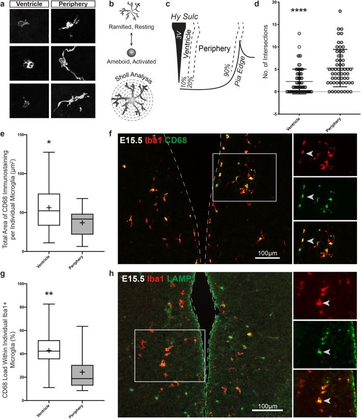
Fig. 3.
Microglia located near the third ventricle are more ameboid at E15.5. a Example phenotypes of E15.5 microglia located near the ventricle and in the periphery. b Schematic illustration depicting microglia resting and activated phenotypes and how the concept of Sholl analysis is applied for analyzing microglia processes. c Illustration defining the ventricle and periphery area in the tuberal hypothalamus used in the microglia Sholl analysis and CD68 analysis. Ventricle microglia were defined as being localized within a 10% relative distance away from the ventricle whereas peripheral microglia were located at a 30% or further relative distance from the ventricle. d Sholl analysis comparing the activation state of E15.5 microglia located near the ventricle (mean value 2.3 ± 2.8 intersections) and in the periphery (mean value 5.3 ± 4.2 intersections). Scatter plot points display individual microglia scores (microglia cells counted within three brain sections from three separate embryos, n = 69 cells in ventricle, n = 59 cells in periphery; P < 0.0001). e Total area of CD68 within microglia cells located near the ventricle (mean value = 56.28 ± 30.16 μm2) and in the periphery (mean value = 36.91 ± 19.67 μm2). f Representative image of microglia activation marker, CD68, and Iba1 at E15.5. Left image displays CD68 and Iba1 expression in the tuberal hypothalamus. Right images are of the boxed area near the ventricle showing CD68 co-labeling with Iba1. Arrow points to example of a co-labeled cell. g CD68 load within microglia cells located near the ventricle (mean value = 42.86 ± 17.60%) and in the periphery (mean value = 24.23 ± 14.77%; 5 microglia cells analyzed from each area in a rostral tuberal hypothalamic brain section from three separate embryos, n = 15 cells in ventricle, 15 cells in periphery; P = 0.0464 for CD68 total area and P = 0.0039 for CD68 load). h Representative image of the lysosomal marker, LAMP-1, and Iba1 at E15.5. Left image shows several Iba1+ microglia co-label with LAMP-1 near the third ventricle. Right images are higher magnifications of boxed area highlighting the co-labeling of Iba1 and LAMP-1. Arrow points to example of a co-labeled cell. Graphs: Scatter plot with mean ± SD. Box and Whisker plots with middle line representing median, cross representing mean, box extending from the 25th to 75th percentiles, and whiskers at max and min values. Statistics: Two-tailed Mann-Whitney U test used in Sholl analysis otherwise Student’s t test, *P ≤ 0.05, **P ≤ 0.01, ****P ≤ 0.0001

