Fig. 6.
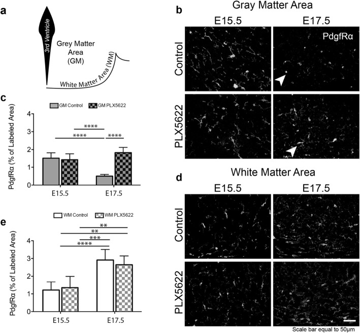
Microglia influence the phenotype of PdgfRα+ late embryonic OPCs in the tuberal hypothalamus. a Illustrative diagram depicting the GM and WM areas from which PdgfRα immunolabeled sample bins were set to capture percent area coverage calculations. b Representative PdgfRα immunostained regions from control and PLX5622 treated embryonic coronal brain slices from the GM area at E15.5 and E17.5. Arrows indicate examples of PdgfRα+ cells that appear amoeboid in the control but ramified in the PLX5622 treated. c Percent area coverage of PdgfRα+ cells from control and PLX5622 treated embryonic coronal brain slices at E15.5 and E17.5 from the GM area (ANOVA, P < 0.0001). d Representative PdgfRα immunostained regions from control and PLX5622 treated embryonic coronal brain slices from the WM area at E15.5 and E17.5. e Percent area coverage of PdgfRα+ cells from control and PLX5622 treated embryonic coronal brain slices at E15.5 and E17.5 from the WM area (ANOVA, P < 0.0001). Samples: n = 5, 5 embryo brains for control and PLX5622 respectively for both E15.5 and E17.5 time points. Graphs: Bar graph with mean ± SD. Statistics: ANOVA with Tukey post hoc; **P ≤ 0.01, ***P ≤ 0.001, ****P ≤ 0.0001

