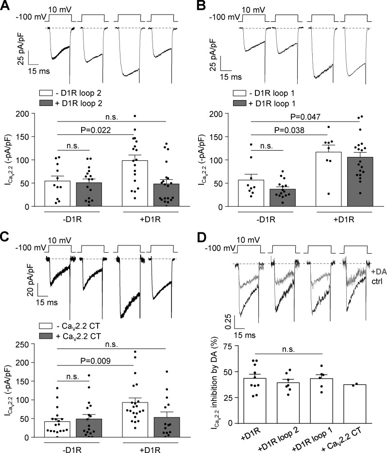
Figure 6.
CaV2.2 current increases caused by D1R constitutive activity are prevented by coexpression of D1R loop 2 and CaV2.2 C terminus without changes in dopamine-mediated CaV2.2 current inhibition in transfected HEK293T cells. (A) Representative traces and averaged CaV2.2 currents (ICav2.2) from HEK293T cells cotransfected with CaV2.2, CaVα2δ1, CaVβ3, and either D1R (+D1R white bar; n = 18) or empty plasmid (−D1R white bar; n = 11), with (+D1R gray bar; n = 20) or without (−D1R gray bar; n = 16) D1R loop 2 (0.2 µg cDNA per well). (B) Representative traces and averaged ICav2.2 from HEK293T cells cotransfected with CaV2.2, CaVα2δ1, CaVβ3, and either D1R (+D1R white bar; n = 8) or empty plasmid (−D1R white bar; n = 9), with (+D1R gray bar; n = 19) or without (−D1R gray bar; n = 15) D1R loop 1 (0.2 µg cDNA per well). (C) Representative traces and averaged CaV2.2 currents (ICav2.2) from HEK293T cells cotransfected with CaV2.2, CaVα2δ1, CaVβ3, and either D1R (+D1R white bar; n = 21) or empty plasmid (−D1R white bar; n = 18), with (+D1R gray bar; n = 14) or without (−D1R gray bar; n = 17) proximal portion of CaV2.2 C terminus containing plasmid (CaV2.2 CT; 0.2 µg cDNA per well). (D) Representative traces and averaged percentage of inhibition of ICav2.2 by dopamine (+DA; 10 µM) in HEK293T cells coexpressing CaV2.2, CaVα2δ1, CaVβ3, and D1R (+D1R; n = 10), D1R plus D1R loop 2 (+loop 2; n = 7), D1R plus D1R loop 1 (+loop 1; n = 6), and D1R plus CaV2.2 CT (+CaV2.2 CT; n = 2). One-way ANOVA with Dunn’s post-test versus −D1R white bar (A and C) and versus +D1R (D); Kruskal-Wallis test with Dunn’s post-test versus −D1R white bar (B). n.s., nonstatistically significant. Data were expressed as mean ± SEM, and dots represent individual data points.

