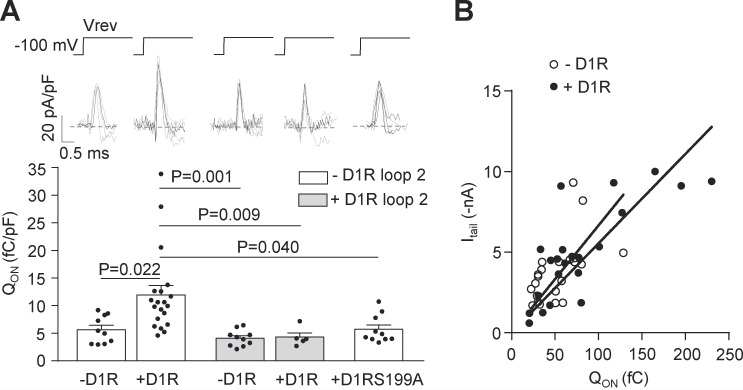
Figure 7.
D1R constitutive activity increases the number of functional CaV2.2 channels at the plasma membrane. (A) Individual (gray) and averaged (black) traces of ON gating CaV2.2 currents (top panel) and averaged QON from HEK293T cells cotransfected with CaV2.2, CaVα2δ1, CaVβ3, and either D1R (+D1R white bar; n = 17), D1RS199A (+D1RS199A white bar; n = 10), or empty plasmid (−D1R white bar; n = 10), with (+D1R gray bar; n = 5) or without (−D1R gray bar; n = 10) D1R loop 2 (0.2 µg cDNA per well; bottom panel). Reversal potential (Vrev, ∼60 mV) was estimated individually for each cell. (B) QON versus peak CaV2.2 tail current (Itail at −60 mV) plot for HEK293T cells coexpressing CaV2.2, CaVα2δ1, CaVβ3 with (+D1R black dots; n = 22, r2 = 0.61) or without D1R (−D1R open dots; n = 22, r2 = 0.31). Lineal regression slopes are not different between the groups (P = 0.187; F = 1.798). One-way ANOVA with Tukey’s post-test (only significant comparisons shown, A). Extra sum of squares F test (B). pF, picofaradays. Data were expressed as mean ± SEM, and dots represent individual data points.

