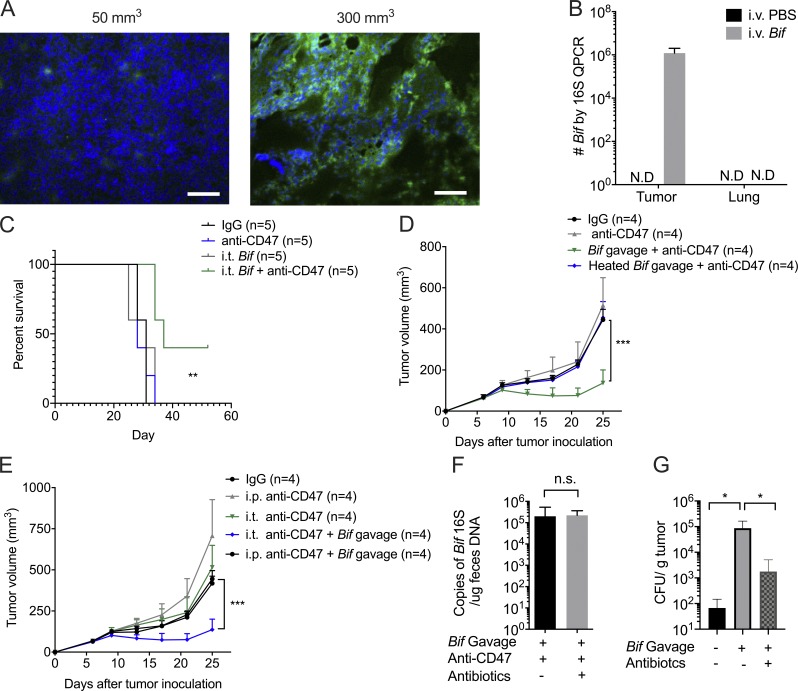
Figure S1.
Gut microbiota could survive in the hypoxic TME and prolong mouse survival. Related to Fig. 2. (A) Representative image of HIF1α stain of different volumes of tumors. Immunofluorescence analysis of hypoxia: HIF1α (green) expression on different-volume tumor tissue slides. Tumor volume of the left: 50 mm3; tumor volume of the right: 300 mm3. Scale bars, 20 µm. One representative experiment is depicted from two experiments yielding similar results. (B) Accumulation of Bifidobacterium inside tumors but not lung sites after treatments. MC38 tumor–bearing Tac mice were i.v. injected with 1.5 × 10⁷ CFU Bifidobacterium or PBS three times when tumor size approaches 200 mm3. 7 d later, tumor tissues were collected for qPCR detection of Bifidobacterium. One representative experiment from two experiments yielding similar results is depicted. N.D., not detected. (C) Survival curves of tumor-burden mice with different treatments. MC38 tumor-bearing Tac mice were i.t. injected with 70 µg anti-CD47 antibody (Ab) or 70 µg rat IgG on days 9 and 13, respectively. For i.t. injection of Bifidobacterium, mice were i.t. injected with 2.5 × 10⁶ CFU Bifidobacterium or PBS on days 6, 9, and 13. Mice were euthanized when the tumor size approached end points. The anti-CD47+ i.t. Bifidobacterium group is compared with the IgG group. One representative experiment from at least two experiments yielding similar results is depicted. **, P < 0.01 (log-rank test). (D) Heat-killed Bifidobacterium failed to facilitate CD47-based immunotherapy. Newly arrived Tac C57BL/6 mice were subcutaneously injected with 5 × 10⁵ MC38 cells. Each mouse was i.t. injected with 70 µg anti-CD47 Ab or 70 µg rat IgG on days 9 and 13, respectively. The mice with Bifidobacterium administration were gavaged with 5 × 10⁸ Bifidobacterium or heat-inactivated Bifidobacterium on days 6, 9, and 13. Tumor volume was measured at the indicated time points. ***, P < 0.001 (two-way ANOVA). (E) Oral administration of Bifidobacterium failed to facilitate antitumor efficacy after systemic administration of anti-CD47 antibody. Newly arrived Tac C57BL/6 mice were subcutaneously injected with 5 × 10⁵ MC38 cells. Each mouse was i.t. injected with 70 µg anti-CD47 Ab or 70 µg rat IgG on days 9 and 13 or i.p. injected with 500 µg anti-CD47 or rat IgG on days 9 and 13, respectively. The mice with Bifidobacterium administration were gavaged with 5 × 10⁸ CFU Bifidobacterium on days 6, 9, and 13, respectively. Tumor volume was measured at indicated time points. ***, P < 0.001 (two-way ANOVA). (F) I.t. antibiotics therapy does not reduce the Bifidobacterium copies in mouse feces. qPCR detection of Bifidobacterium in fresh feces collected from mice with indicated treatments on day 14 (also see Fig. 2 H; nonpaired Student’s t test). (G) Detection of Bifidobacterium inside tumor tissues after Bifidobacterium gavage and i.t. antibiotics treatment. Newly arrived Tac C57BL/6 mice were subcutaneously injected with 5 × 10⁵ MC38 cells. For oral administration of Bifidobacterium (Bif), mice were gavaged with 5 × 10⁸ CFU Bifidobacterium or PBS on day 7, 10, and 13. Mice were i.t. injected with 100 µl antibiotic cocktail solution or PBS every other day from day 6. Tumor tissues were collected on day 17 and homogenized for anaerobic culture of Bifidobacterium. Presented as mean ± SEM. *, P < 0.05 (Mann–Whitney U test of log scale). n.s., not significant.

