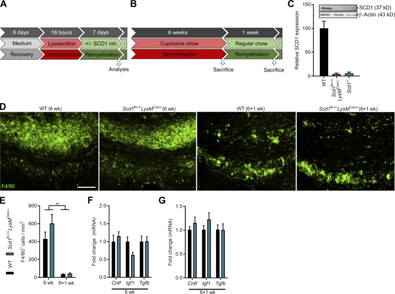
Figure S4.
Confirmation of SCD1 ablation and pathological parameters of Scd1fl+/+LysmCre+ mice. (A and B) Experimental setup of the brain slice culture model for remyelination (A) and cuprizone-induced acute demyelination in vivo model (B). SCD1 inh., SCD1 inhibitor. (C) Immunoblot analysis of SCD1 protein in WT, Scd1fl+/+LysmCre+/−, and Scd1−/− BMDMs stimulated with the LXR agonist T0901317 (n = 2 cultures of different animals). (D and E) Representative immunofluorescence images and quantification of F4/80 staining of CC from WT (6 wk, n = 11 animals; 6+1 wk, n = 10 animals) and Scd1fl+/+LysMCre+/− mice (6 wk, n = 7 animals; 6+1 wk, n = 6 animals). Scale bar, 100 µm. (F and G) mRNA expression of neurotrophic factors in CC of WT (6 w, n = 11 animals; 6+1 wk, n = 10 animals) and Scd1fl+/+LysMCre+/− mice (6 w, n = 7 animals; 6+1 wk, n = 6 animals) after cuprizone-induced demyelination (6 w) and subsequent remyelination (6+1 wk). All replicates were biologically independent. All data are represented as mean ± SEM. **, P < 0.01, unpaired Student’s t test (E–G).

