Fig. 3.
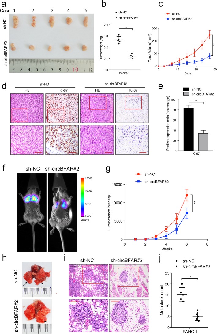
CircBFAR promotes tumor growth and metastasis of PDAC cells in vivo. a Representative images of subcutaneous xenograft tumors. b, c The tumor volume and weight dramatically decreased in sh-circBFAR#2 treated mice compared with those treated with the control shRNA. d, e Representative HE and IHC staining images of subcutaneous tumors revealed the relative protein levels of Ki-67 in different groups. The images were photographed at 200X (upper panel) or 400X (lower panel) magnification. Scale bar: black =100 μm; red =50 μm. f, g Representative IVIS images and analysis of luminescence intensity in lung in tail-vein injection model (n = 6 for each group). h Representative images of lung metastatic tumors. i HE staining of lung metastatic tumors. The images were photographed at 100X (upper panel) or 200X (lower panel) magnification. Scale bar: black =200 μm; red =100 μm. j The number of lung metastatic tumors decreased significantly in sh-circBFAR#2 treated mice. Statistical significance was assessed using two-tailed t-tests for two group comparison, and one-way ANOVA followed by Dunnett’s tests for multiple comparison. The error bars represent the standard deviations of three independent experiments. **P < 0.01

