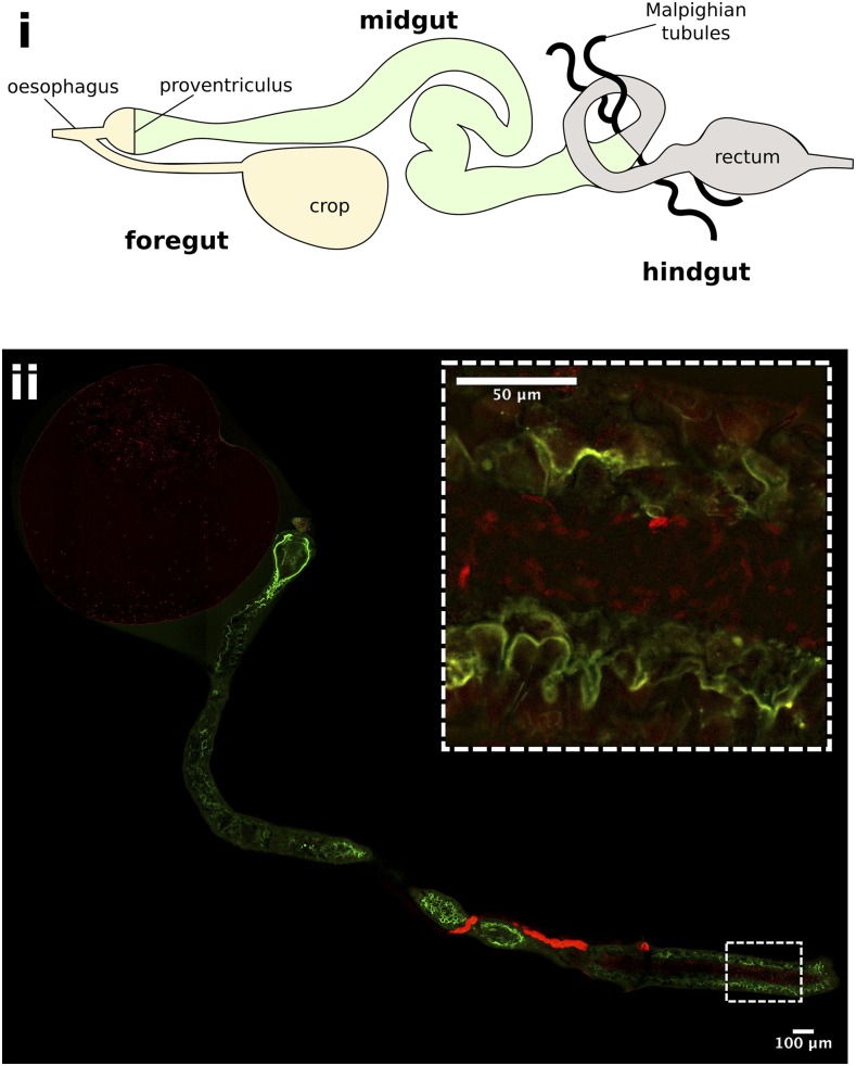
Figure 3.
Herpetomonas muscarum in the fly digestive tract immediately after feeding. i - Schematic of the D. melanogaster digestive tract. ii – The foregut and midgut of D. melanogaster two hours after feeding with tdTomato expressing H. muscarum. The flies used for this experiment express a myosin-GFP fusion protein to allow the gut epithelial border to be visualized. H. muscarum can be seen in the crop and the midgut (inset) of the fly.

