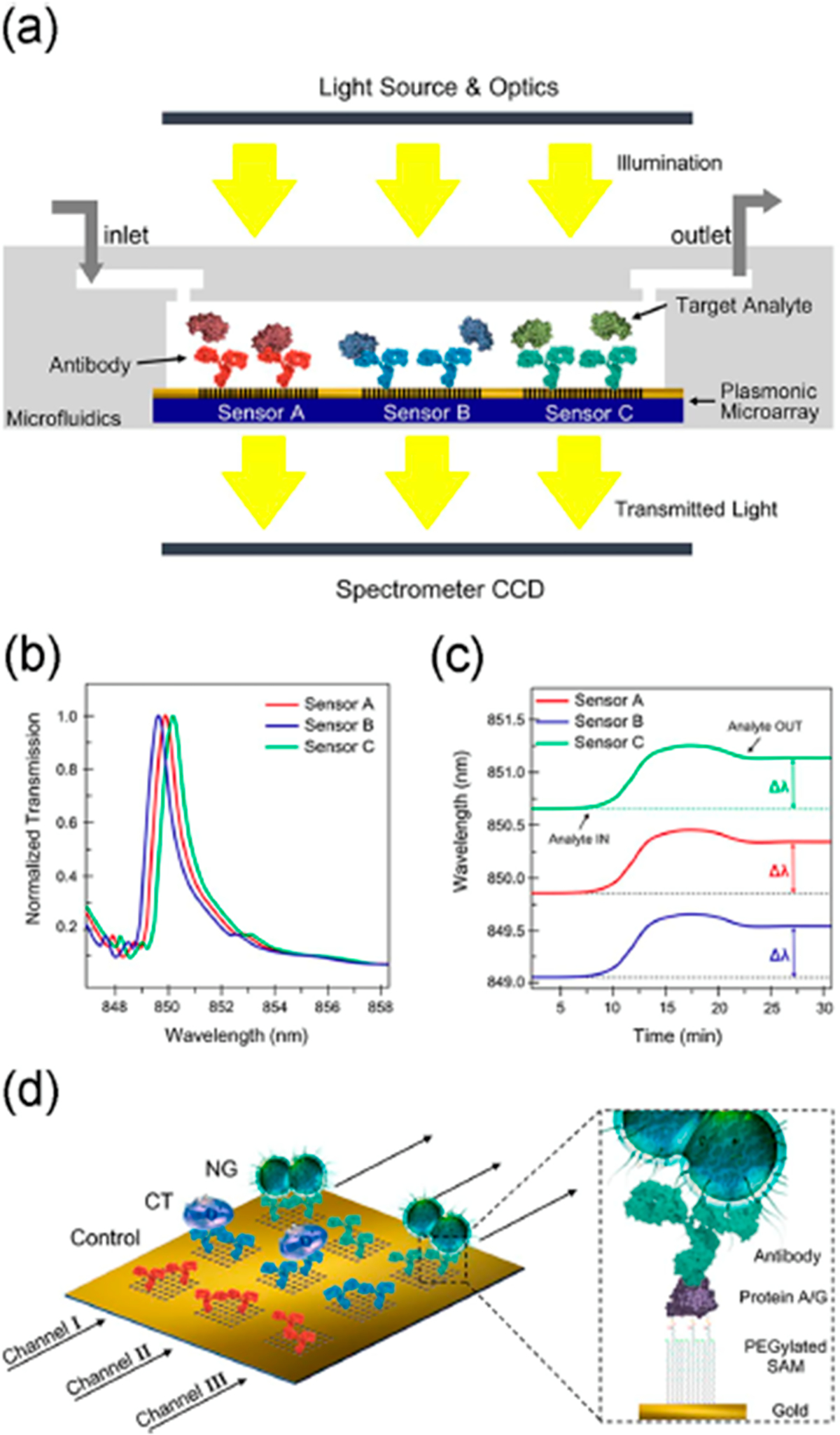
Figure 7.

Nanohole biosensor: (a) cross-sectional view, (b) graph illustrating the three EOT spectral peaks acquired simultaneously from the three sensor arrays, (c) graph illustrating the multiplexed real-time monitoring of the EOT wavelength shift, and (d) schematics of sensor surface biofunctionalization. Different nanohole arrays are decorated with different antibodies: anti-Neisseria gonorrheae (NG) (green), anti-Chlamydia trachomatis (CT) (blue), and a control antibody (red). Each microfluidic channel (black arrows) cover three inline sensor arrays. The zoomed-in section illustrates the surface chemistry for the sensor functionalization. Reprinted from Biosensors and Bioelectronics, Vol. 94, Sole, M.; Belushkin, A.; Cavallini, A.; Kebbi-Beghdadi, C.; Greub, G.; Altug, H. Multiplexed nanoplasmonic biosensor for one-step simultaneous detection of Chlamydia trachomatis and Neisseria gonorrheae in urine, pp. 560–567 (ref 69). Copyright 2017, with permission from Elsevier.
