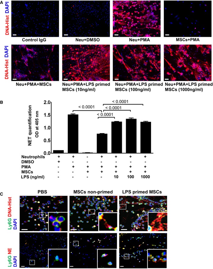
Figure 1. LPS‐primed MSCs activate neutrophils with enhanced NET .

- MSCs were exposed to increasing LPS concentrations, thereafter washed and co‐cultured with PMA‐activated neutrophils. Incubation of neutrophils with PMA alone served as a positive control and DMSO‐treated neutrophils as a negative control. Scale bars: 50 μm.
- Quantification of NET‐bound elastase indicative of NET formation. Statistical analysis was performed using one‐way ANOVA, and values are represented as mean ± SEM, n = 3 biological replicates.
- Representative microphotographs of murine wound sections immunostained for Ly6G (neutrophils, green) and DNA‐histone (displayed as expulsed streaks in red indicative of NETs). Wounds injected with LPS‐primed MSCs (upper row, outer right panel) depict enhanced NET formation (magnified inset) and increased expression of activation markers like neutrophil elastase (NE, lower panel, outer left panel) compared to wounds injected with non‐primed MSC (middle panels) or PBS‐injected wounds (outer left panels). Murine skin wounds treated with PBS injections served as control. Scale bar: 10 μm (upper row) and 50 μm (lower row). To facilitate comparison, areas inside the rectangles are shown at 5× magnification in the insets.
Source data are available online for this figure.
