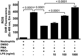
Figure EV1. LPS‐treated MSCs augment neutrophil activation and enhanced ROS production.

Graph shows quantification of ROS by measuring fluorescence intensity of DHR123 dye. The MSCs were primed with increasing concentrations of 10 ng/ml, 100 ng/ml, and 1,000 ng/ml LPS followed by co‐culture with neutrophils. The MSCs were then incubated with ROS‐specific dye DHR123, and the fluorescence was read after 60 min of incubation at 488/520 nm with spectrophotometer. Incubation of neutrophils with PMA alone served as a positive control and DMSO‐treated neutrophils as a negative control. Statistical analysis was performed using one‐way ANOVA, and values are represented as mean ± SEM, three biological replicates.
