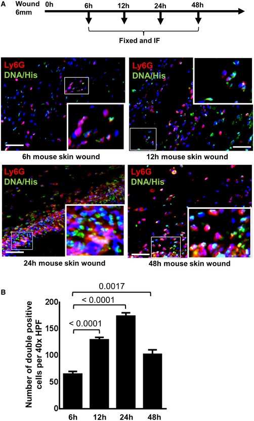
Figure EV2. Recruitment of neutrophils increases with time after induction of wounds in mice.

- Immunostaining of Ly6G (green) and DNA‐histone (DNA‐Hist, red) was performed at 6, 12, 24, and 48 h after induction of full‐thickness wounds. Nuclei were stained by DAPI (blue). Staining with Ly6G for neutrophils and DNA‐histone for NET formation allowed to study neutrophil recruitment and NET formation. To facilitate comparison, areas inside the rectangles are shown at 5× magnification in the insets. Scale bars: 50 μm
- Quantitative analysis of Ly6G and DNA‐histone double‐positive cells at different time intervals. Statistical analysis was performed using one‐way ANOVA, and values are represented as mean ± SEM, six biological replicates.
