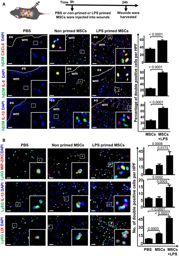
Figure 3. LPS‐primed MSCs increase expression of neutrophil regulatory genes in vivo .

- The upper scheme depicts the experimental design. Representative microphotographs show that injection of LPS‐primed MSCs into wounds increased the expression of CXCL6, IL‐8, and IL‐1β. Graphs display numbers of double‐positive cells for h‐β2M + CXCL6, h‐β2M + IL‐8, and h‐β2M + IL‐1β, respectively. MSC‐injected wounds served as controls. Statistical analysis was performed using unpaired t‐test, and values are represented as mean ± SEM, six biological replicates. To facilitate comparison, areas inside the rectangles are shown at 5× magnification in the insets. Es, eschar on wound margin; wm, wound margin; scale bars: 50 μm.
- Results show that LPS‐primed MSCs injected into wounds provoke increased expression of MIP/KC in endogenous neutrophils. MIP/KC represents functional homologues of IL‐8 in mice and is known as neutrophil chemoattractant. Similar results were found for IL‐1β, which is a strong chemoattractant for neutrophil recruitment and bacterial clearance. In addition, LIX which shares 63% amino acid sequence identity with human GCP‐2/CXCL6, a chemoattractant for neutrophils. The graphs display numbers of double‐positive cells for Ly6G MIP/KC, IL‐1β, and LIX, respectively. PBS and non‐primed MSC‐injected wounds served as controls. To facilitate comparison, areas inside the rectangles are shown at 5× magnification in the insets. Statistical analysis was performed using one‐way ANOVA, and values are represented as mean ± SEM, six biological replicates. Scale bars: 50 μm.
