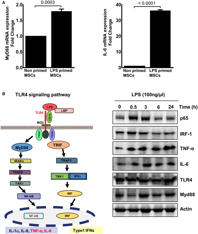
Figure EV3. LPS‐treated MSCs activate MyD88 and IL‐6 expression in the TLR4 signaling pathway.

- Human MSCs were treated with LPS (100 ng/ml) for 24 h. The mRNA expression of MyD88 and IL‐6 was assessed by qRT–PCR analysis at 24 h. MyD88 (upper left panel) and IL‐6 gene expression (upper right panel) revealed an increase in MyD88 and IL‐6 in LPS‐primed MSCs. Statistical analysis was performed using unpaired t‐test, and values are represented as mean ± SEM, three biological replicates.
- The TLR4 signaling pathway is depicted. Upon LPS binding, conformational changes in the TLR4 receptor led to recruitment of intracellular TLR domains harboring adaptor proteins. A bifurcation in MyD88‐dependent signaling converges to NF‐κB and pro‐inflammatory cytokine release, among them IL‐6, and the TRIF signaling pathway relaying its signals to downstream effectors like the transcription factor IRF which transactivates type 1 interferons. The right panel depicts representative Western blot analyses with a time‐dependent regulation of TLR4 and components of the TLR4 signaling pathway after exposure of MSCs with LPS. p65, a component of the heterodimeric NF‐κB transcription factor, and the IRF‐1 transcription factor are swiftly induced at 0.5 h, but down‐regulated to basal levels at 6 h after LPS challenge of MSCs. The target genes of NF‐κB are induced at 0.5 h, and this induction is differentially maintained. The expression of indicated proteins was analyzed in the cell lysates from LPS (100 ng/ml)‐ or PBS‐treated MSCs, three biological replicates.
Source data are available online for this figure.
