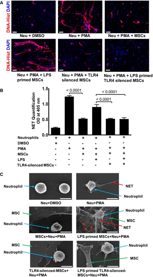
Figure 4. TLR4‐silenced MSCs fail to activate neutrophils and to enhance NET formation.

- Representative microphotographs of TLR4‐silenced and non‐silenced MSCs primed with LPS and co‐cultured with neutrophils. Immunostaining was performed with an antibody detecting NETs (DNA‐histone, red). Of note, TLR4‐silenced MSCs upon LPS priming cannot mount any adaptive response and fail to enhance NET formation (lower row, outer right panel) as compared to non‐silenced, LPS‐primed MSCs co‐cultured with PMA‐activated neutrophils (lower row, outer left panel). Neutrophils and DMSO served as a negative control (upper row, outer left panel), while neutrophils activated by the protein C kinase activator PMA served as a positive control depicting enhanced NET formation (upper row, middle panel). Co‐culture with MSCs suppressed enhanced NET formation (upper row, outer left panel) induced by PMA. Scale bars: 50 μm.
- Quantitative assessment of NET‐bound elastase employing a specific ELISA in identical experimental setting as described in Fig 1. Similarly, to immunostaining, a significant reduction of NET‐bound elastase was observed in TLR4‐silenced LPS‐primed MSCs when co‐cultured with activated neutrophils as opposed to high NET‐bound elastase in non‐silenced LPS‐primed MSCs co‐cultured with activated neutrophils. Statistical analysis was performed using one‐way ANOVA, and values are represented as mean ± SEM, three biological replicates.
- High‐resolution scanning electron microscope analysis depicting enhanced NET formation (red arrows) expulsed from neutrophils (blue arrows) in the presence of LPS‐primed MSC co‐cultured with PMA‐activated neutrophils (middle row, outer left panel) as compared to reduced NETs in non‐primed MSCs co‐cultured with activated neutrophils (middle row, outer right panel). Of note, TLR4‐silenced LPS‐primed MSCs failed to activate neutrophils and NETs (lower row, outer left panel). TLR4‐silenced and non‐primed MSCs display reduced NETs in co‐cultures with activated neutrophils (lower row, outer right panel), suggesting that PMA activation shares TLR4 signaling components. PMA‐activated neutrophils alone served as a positive control with highly enhanced NETs (upper row, outer right panel) and DMSO‐treated neutrophils as negative controls (upper row, outer left panel). Red stars indicate neutrophil‐derived granules. Scale bars: 0.1 μm
